
T Cell Receptors
By Jennifer
McDowall
![]()
To view structure of T cell receptors
|
|
|
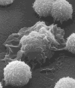
Scanning electron micrograph of a single macrophage surrounded by
several lymphocytes. Courtesy of
J.M. Orenstein, George Washington University, and from ‘Kimball’s
Biology Pages’ at: |
Your blood is composed of different types of cells, one of which is the white blood cell, or leukocyte, that forms part of your body’s defence system against intruders such as viruses, bacteria, and parasites. Lymphocytes, monocytes and granulocytes are all types of white blood cells with specialised roles to play. Of these, the lymphocytes are critical for mounting an adaptive immune response, which specifically targets the invading pathogen. The B cell lymphocytes are responsible for a humoral adaptive response, which involves the production of highly specific antibodies, while T cell lymphocytes are responsible for a variety of cell-mediated adaptive responses. These responses include the destruction of viral-infected or malignant cells by cytotoxic T lymphocytes, enhancing the B cell antibody response by helper T cells, and the recruitment of macrophages and other cells to the site of infection by inflammatory T cells. In each case, the B or T cell is specific for a particular antigen, but they differ in how they detect those antigens. The receptors on the surface of B cells (BCRs) can bind to soluble antigens, while T cell receptors (TCRs) can only recognise an antigen when it is complexed with major histocompatibility complex (MHC) molecules on the surface of other cells.
T Cell
Receptors, Triggering a Response
|
|
|
T cell receptor binding to MHC-antigen complex. Courtesy of J. Kimball,
from: |
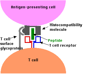
T cell receptors recognise foreign antigens, then convey the message to the nucleus to induce a response. The body produces many T cells, each with specific TCRs on its surface through the recombination of the genes that encode the receptors, before it has encountered complementary antigens. By having a surplus of T cells carrying different TCRs, the body is able to mount a fast response once an intruder is detected. Furthermore, there are thousands of identical TCRs on the surface of a T cell, which increases the likelihood of binding when an antigen is encountered. The antigens that TCRs bind are small peptide fragments, or epitopes, displayed by MHC molecules on the surface of cells. Cytotoxic T lymphocyte TCRs recognise epitopes displayed by MHC class I molecules on the surface of almost every cell in the body, so it can distinguish between ‘self’-antigens and foreign antigens (viral-infected cells), as well as being sensitive to the amount of self-antigen presented (increased number of self-antigens in malignant cells). Helper T cell and inflammatory T cell TCRs recognise epitopes displayed by MHC class II molecules on the surface of antigen-presenting immune cells, including: macrophages that engulf foreign particles such as bacteria; dendritic cells that present antigen to T cells; and B cells that produce antibodies. The binding of the epitope to the TCR involves a T cell surface glycoprotein: CD8 on cytotoxic T lymphocytes, and CD4 on helper T cells and inflammatory T cells (as shown in the picture to the left). The CD8 and CD4 surface glycoproteins recognise MHC class I and II molecules, respectively. The binding of a TCR to an epitope can result in a signal being sent to the nucleus to induce a response.