
Snake Venom:
Bungarotoxins
By
Jennifer McDowall
![]()
To view acetylcholinesterase structure
Venom: an arsenal of toxins
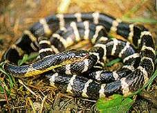
Many-banded
krait
 Venomous
snakes are found throughout the world; they are even lurking in our
oceans. Said to cause over 3 million
bites a year worldwide, they pose a significant health problem both in
mortality and morbidity, causing amputations and renal failure, in addition to
over 100,000 deaths a year. The major
culprits are vipers, elapids, colubrids and sea snakes. Of these, elapids such as cobras and kraits
have developed the most potent toxins.
Snake venoms contain a multitude of biologically active toxins that work
together for the capture of prey (take a look at the Table
listing the
variety of toxins found in the venom of the many-banded krait). Their effects include pro- and anti-blood
coagulation, neurotoxicity, mycotoxicity, nephrotoxicity, cardiotoxicity and
necrotoxicity (local tissue damage). Amongst these, the neurotoxins play a key
role in immobilising prey through paralysis, disorientation and depressed
respiration.
Venomous
snakes are found throughout the world; they are even lurking in our
oceans. Said to cause over 3 million
bites a year worldwide, they pose a significant health problem both in
mortality and morbidity, causing amputations and renal failure, in addition to
over 100,000 deaths a year. The major
culprits are vipers, elapids, colubrids and sea snakes. Of these, elapids such as cobras and kraits
have developed the most potent toxins.
Snake venoms contain a multitude of biologically active toxins that work
together for the capture of prey (take a look at the Table
listing the
variety of toxins found in the venom of the many-banded krait). Their effects include pro- and anti-blood
coagulation, neurotoxicity, mycotoxicity, nephrotoxicity, cardiotoxicity and
necrotoxicity (local tissue damage). Amongst these, the neurotoxins play a key
role in immobilising prey through paralysis, disorientation and depressed
respiration.
 Venoms often contain different
neurotoxins that work synergistically to cripple the nervous system. Neurotoxins can be classified according to
their site of action: pre-synaptic neurotoxins block neurotransmission by
affecting acetylcholine transmitter release; post-synaptic neurotoxins are
antagonists of the acetylcholine receptor.
Together these neurotoxins effectively block skeletal neuromuscular
transmission by crippling receptors, while at the same time acting to destroy
any neurotransmitter that might compete with the toxin for receptor binding. Venoms often contain several post-synaptic
neurotoxins, each with a high affinity for a nicotinic receptor subtype - in
this way the venom can cripple as many receptors as possible. The post-synaptic neurotoxins are found only
in elapids and sea snakes (Hydrophiidae). In the many-banded krait pictured above, a pre-synaptic toxin is b-bungarotoxin, while post-synaptic toxins
are a‑ and k-bungarotoxins.
Venoms often contain different
neurotoxins that work synergistically to cripple the nervous system. Neurotoxins can be classified according to
their site of action: pre-synaptic neurotoxins block neurotransmission by
affecting acetylcholine transmitter release; post-synaptic neurotoxins are
antagonists of the acetylcholine receptor.
Together these neurotoxins effectively block skeletal neuromuscular
transmission by crippling receptors, while at the same time acting to destroy
any neurotransmitter that might compete with the toxin for receptor binding. Venoms often contain several post-synaptic
neurotoxins, each with a high affinity for a nicotinic receptor subtype - in
this way the venom can cripple as many receptors as possible. The post-synaptic neurotoxins are found only
in elapids and sea snakes (Hydrophiidae). In the many-banded krait pictured above, a pre-synaptic toxin is b-bungarotoxin, while post-synaptic toxins
are a‑ and k-bungarotoxins.

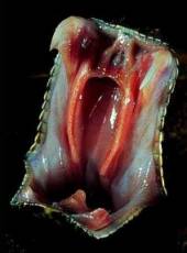
Milking a snake
Photographs courtesy of B. G. Fry, Australian Venom Research Unit, Melbourne, Australia
Next: The neuromuscular junction