
Haemoglobin
Family Ties
Haemoglobin is part of the large globin family of haem-containing proteins that are involved in the binding and/or transport of oxygen. These proteins are widely distributed in many organisms. Examples of some other globin proteins found in this family are:
Ø Myoglobin (used as a reserve supply of oxygen, and facilitates the movement of oxygen within muscles)
Ø Neuroglobin (involved in oxygen transport in the brain)
Ø Cytoglobin (involved in intracellular oxygen storage or transfer)
Ø Leghaemoglobin (provides oxygen to bacteroids, which is essential for symbiotic nitrogen fixation)
Ø Erythrocruorin (giant haemoglobin of worms)
Ø Plant haemoglobin (may act as an oxygen sensor)
Ø Flavohaemoglobin (involved in NO detoxification)
All of these proteins share a common alpha-helical globin structure.
What InterPro Tells Us
As the same signatures hit many of the globins, only the beta globin chain will be used as an example:
P68871 Human Beta Globin Chain
InterPro Domain Architecture
![]()
InterPro Entry |
Method Accession |
Graphical Match |
Method Name |
|
IPR000971 |
PF00042 |
|
globin |
|
IPR000971 |
PS01033 |
|
GLOBIN |
|
IPR002337 |
PR00814 |
|
BETAHAEM |
|
IPR009050 |
SSF46458 |
|
Globin_like |
|
IPR012292 |
G3D.1.10.490.10 |
|
Globin_related |
|
Classification |
PDB Chain/Domain ID |
PDB Chain/Structural Domains |
|
1ird
|
1irdB
|
|
|
|
1.10.490.10.4 |
1irdB0 |
|
|
|
a.1.1.2 |
d1irdb_ |
|
|
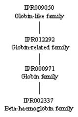
From the graphical match above, you can see that the
signatures (method accession) are grouped into four InterPro entries for the human
alpha globin chain. These reflect hierarchical
relationships between the different signatures. IPR009050, the
globin-like family, is top of the hierarchy and has one signature, SSF46458 from the SUPERFMAILY
database. This entry represents any
protein with a globin-like structure that is evolutionarily related to globin,
which includes the entire globin family, as well as truncated haemoglobins and phycocyanin-like
phycobilisome proteins. The entry IPR012292, the
globin-related family, represents proteins more closely related in sequence and
structure than those found in IPR009050 (lacks the more distantly related phycocyanin-like
phycobilisome proteins), and has one signature: G3D.1.10.490.10 from the
Gene3D database. The next entry is IPR000971, the globin family,
that represents the globin family itself (closely related in sequence), and has
two signatures: PF00042 from the PFAM database and
PS01033 from the PROSITE
database. The remaining entry, IPR002337, the
beta-haemoglobin family, is the most specific classification, and is
represented by one signature: PR00814 from the
PRINTS database. As all five of these
signatures cover the same sequence, they are related to one another: IPR009050 is the parent of IPR012292,
which in turn is the parent of IPR000971, which is itself the parent of
IPR002337:
The
remaining three entries in the table above are from the structural database PDB
(green stripe), and from the structural classification databases CATH (pink
stripe) and SCOP (black stripe) (the names such as d1irdb_ being derived from the PDB entry
upon which it is based, here PDB entry 1ird, chain B). The graphical match for the PDB entry 1ird displays the length of the original PDB entry, which covers the
entire protein. The CATH (1irdB0) and SCOP (d1irdb_ ) databases give information on
the classification of this protein.
What the Structure Tells Us
Haemoglobin
was the first protein to have its 3-dimensional structure determined by Max
Perutz and his colleagues at Cambridge University in the 1950s, and has served
as a model to correlate a protein’s structure with its function. The structures of the different globin chains
can be viewed through InterPro using AstexViewer®, which is linked from the
InterPro Match Table of each protein via the logo ![]() (please
note, there is no link directly from this page to the AstexViewer® for the
protein discussed above, therefore you need to go to the link on the InterPro
pages for P68871). The AstexViewer® displays the PDB structure
with the particular CATH or SCOP domain highlighted in yellow. There are several structures associated the
different haemoglobin chains in the Protein Data Bank (PDB). A detailed description and visualisation of
the structural features of haemoglobin can be found at the PDB ‘Molecule of the
Month’, providing insights into the molecular basis of action for
this oxygen carrier.
(please
note, there is no link directly from this page to the AstexViewer® for the
protein discussed above, therefore you need to go to the link on the InterPro
pages for P68871). The AstexViewer® displays the PDB structure
with the particular CATH or SCOP domain highlighted in yellow. There are several structures associated the
different haemoglobin chains in the Protein Data Bank (PDB). A detailed description and visualisation of
the structural features of haemoglobin can be found at the PDB ‘Molecule of the
Month’, providing insights into the molecular basis of action for
this oxygen carrier.
Next: Table of Haemoglobin and Related Proteins
Previous: Haemoglobin and Disease