Sm Ribonucleoproteins
What InterPro Tells Us:
P14678 Human Sm
ribonucleoprotein B/B’
InterPro Domain Architecture:
![]()
|
InterPro Entry |
Method Accession |
Graphical Match |
Method Name |
|
IPR001163 |
PF01423 |
|
LSM |
|
IPR001163 |
SM00651 |
|
Sm |
|
IPR006649 |
PD020287 |
|
snRNP |
|
IPR010920 |
SSF50182 |
|
Sm_like_riboprot |
|
Classification |
PDB Chain/Domain ID |
PDB Chain/Structural Domains |
|
|
1d3b |
1d3bb |
|
|
|
1d3b |
1d3bd |
|
|
|
1d3b |
1d3bf |
|
|
|
1d3b |
1d3bh |
|
|
|
1d3b |
1d3bj |
|
|
|
1d3b |
1d3bl |
|
|
|
2.30.30.100.4 |
1d3bL0 |
|
|
|
b.38.1.1
|
d1d3bb_
|
|
|
From the graphical match above, you can see that the signatures (method accession) are divided into three InterPro entries for human Sm ribonucleoprotein B/B’. All the signatures above represent the core domain of the Sm ribonucleoprotein B/B’. These signatures also give information about the hierarchy of relationships that involve this protein.
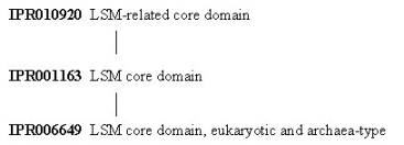
The InterPro entry IPR006649 has one signature representing the core of eukaryotic and archaea-type Lsm ribonucleoproteins: PD020287 from the PRODOM database. IPR006649 represents a subset of IPR001163, which is termed a ‘parent’ entry of IPR006649, and which represents the core of both Lsm ribonucleoproteins and the bacterial Lsm pleiotropic translational factor Hfq, these proteins showing a high degree of sequence identity with each other over this region. Two signatures represent IPR001163: PF01423 from the PFAM database, and SM00651 from the SMART database. IPR001163 is in turn a child of the parent entry IPR010920, which represents proteins containing regions related to the Lsm core. This includes Lsm proteins, bacterial Lsm Hfq proteins, and the middle domain of the mechanosensitive channel of small conductance protein MscS, all of which share a similar SH3-like open beta-barrel topology. IPR010920 is represented by one signature: SSF50182 from the SUPERFAMILY database.
The
remaining eight entries in the table above are from the structural database PDB (green stripe), and from the
structural classification databases CATH (pink stripe) and SCOP (black stripe)
(the names such as d1d3bb_ are derived from the PDB entry upon which they are
based, here PDB entry 1d3b, chain b).
The graphical match for the PDB entry 1d3b displays the length of the
original PDB entry, which as shown here consists of 6 identical chains.
The CATH (1d3bL0) and SCOP (d1d3bb_) databases give information on
the classification of this core domain.
What the Structure Tells Us
Structures
associated with the Sm ribonucleoprotein B/B’ can be viewed using AstexViewer®,
which is linked from the Match Table via the logo ![]() on the
InterPro page (please note, there is no link directly from this page to the
AstexViewer®, therefore you need to go to the link on the InterPro page for P14678).
The AstexViewer® displays the PDB structure with the particular CATH or
SCOP domain highlighted in yellow. There
are many structures associated with Sm ribonucleoprotein B/B’, as well as for
other LSM proteins, in the
Protein Data Bank (PDB).
on the
InterPro page (please note, there is no link directly from this page to the
AstexViewer®, therefore you need to go to the link on the InterPro page for P14678).
The AstexViewer® displays the PDB structure with the particular CATH or
SCOP domain highlighted in yellow. There
are many structures associated with Sm ribonucleoprotein B/B’, as well as for
other LSM proteins, in the
Protein Data Bank (PDB).
Other molecules involved in RNA processing include ribozymes, which are RNA molecules capable of catalysing the cleavage and formation of covalent bonds in RNA strands. A detailed description and visualisation of the structural features of ribozymes can be found at the PDB ‘Molecule of the Month’.
Next: Table of Sm
ribonucleoproteins
Previous: Sm Core Assembly and
Specificity